
During pregnancy, one of the biggest concerns of any couple is that everything goes well and the baby is totally healthy.
Although we cannot absolutely rule out all possible alterations, we have several tests to perform in the first months of pregnancy and rule out most.
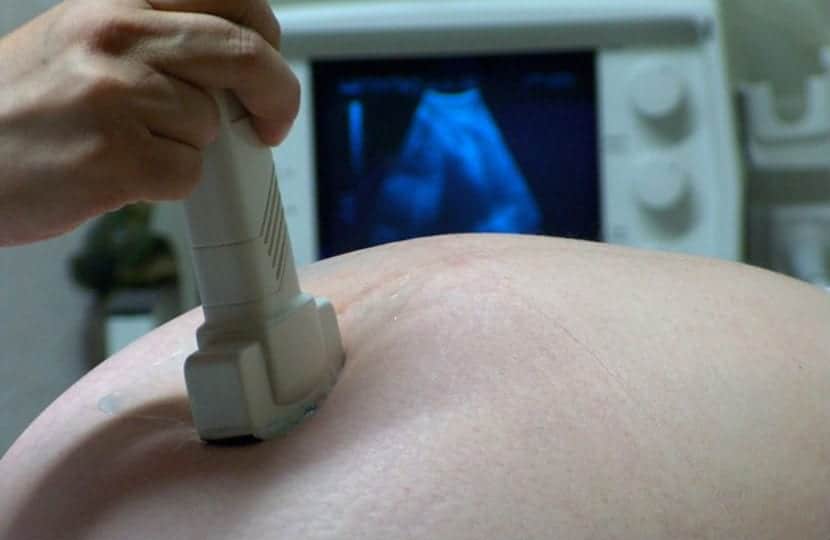
When we reach the 12th week of pregnancy, it is time to perform the first major ultrasound, from this moment on we will be able to perform a series of serial tests, to rule out chromosomal alterations in the baby.
In most cases, The more dangerous invasive tests are not carried out, unless the non-invasive tests raise suspicions of problems.
We are going to see the tests from less to more decisive and also invasive and dangerous.
triple screening
It is, along with the week 12 ultrasound, the first major test that they will do to us in pregnancy regarding the possibility of chromosomal alterations.
A blood draw is made to the mother, in which two hormones are determined, one of them produced by the placenta (PAPPA) and the other, the pregnancy hormone (BHCG) produced at that time by the ovary.
The values of these hormones are combined with the ultrasound data; size of the nuchal fold and presence of nasal bone in the baby.
The mother's age is also taken into account and corrections are made according to the mother's weight and if she is a smoker..
These data are analyzed with a computer program that will give us a statistical risk that our baby is a carrier of Down and Edwards Syndrome.
The result is classified as Low Risk, Intermediate Risk and High Risk. In the event that the risk is low, the parents are not offered further tests. The error rate is 5%.

Fetal DNA test in maternal blood
It is a technique relatively modern, its great advantage is that it is not invasive, so that it does not put the pregnancy at risk.
It is based on evidence of the presence of fetal DNA in the pregnant woman's blood.
A blood sample is taken from the mother and the fetal DNA is detected, then a reconstruction and analysis of that DNA is carried out to detect alterations in the chromosomes.
It is very reliable, but not all chromosomes are analyzed, generally only those that are most frequently altered.
When no alterations are detected, further tests are not recommended. but if the result is altered and a chromosomal problem is suspected, the next test is carried out.

Amniocentesis
It would be the following test to be carried out in case risk appears in any of the previous tests.
It is a invasive test y poses a real risk of pregnancy loss.
It is done between weeks 14 to 18 of pregnancy. First, an ultrasound study is performed in which the amount of amniotic fluid, the location of the placenta and the baby's situation will be detected.
Once these parameters have been assessed, an area is located far from the placenta and the fetus, where the amount of amniotic fluid is abundant.
The test is performed under continuous ultrasound control, they will prick us in the abdomen until we reach that area with a good amount of liquid and slowly inhaling, they will extract about 20 ml of liquid, which is the one that will be analyzed later.
Before concluding the test, it is checked that the baby's heart beats correctly.
In the liquid that has been drawn fetal cells are detected and cultured, so that genetic material can be analyzed, that is, of the chromosomes of the fetus and carry out the complete karyotype, looking for alterations in the number or shape of these chromosomes.
The next 48 hours will recommend rest And if your group is Rh negative, it is important that you receive the anti-D vaccine that prevents your body from creating defenses against Rh if the baby is Rh +.
The final result is obtained in about 20 days, although in the first week the existence of alterations begins to be seen.
Although it does not guarantee 100% that our baby is totally healthy the precision regarding the normality or not of the chromosomes is very close.
Risks
It is an invasive test. The most frequent complications the rupture of the bag of waters, bleeding, infection in the amniotic membranes or abortion.

Chorionic biopsy
It consists of obtaining a sample of the chorionic villi to perform the genetic study.
What are chorionic villi?
The chorion is an envelope that surrounds the embryo and that later will give rise to the placenta, The villi are a part of that envelope that are the ones that will give rise to the mechanism of attachment of said placenta. They are also those that absorb nutritional substances to nourish the embryo.
It can be done from week 10 to 14 and it is even more definitive than amniocentesis, since more genetic material is obtained from the fetus.
With this technique, the fetal karyotype can be performed and also familial congenital diseases, such as cystic fibrosis or Huntington's chorea.
It can be done through the cervix or by puncture in the abdomen.
It is also performed with ultrasound control and before the end of the test it is verified that the embryo's heart rate is normal.
It is also important to rest 24 hours after the test and the anti-D vaccination if the mother is RH-.
The final result is also obtained in 20 days, although a special technique is usually performed for a quick result that, in less than a week, guides us on the existence or not of alterations.
The risks are similar to those of amniocentesis; bag rupture, infection, bleeding, or loss of pregnancy.
cordocentesis
Consists of the blood draw directly from the baby's umbilical cord.
It is important to perform an ultrasound in which the specialist not only locates the placenta, the baby and the amount of amniotic fluid, he also has to locate exactly the path of the umbilical cord.
Afterwards, the puncture is performed through the abdomen, always with ultrasound control, until reaching the umbilical cord and blood is drawn from our baby.
The amount of blood that is extracted in a little, about 3-4 ml, to perform the karyotype of the baby and an analysis to know if there is anemia or another alteration in the baby's blood.
The results are very fast and in about three days we will know if there is any genetic alteration, as well as if there is anemia or any blood disorder ...
It is done later, never before the 18th week of pregnancy. It is performed when ultrasound scans (especially at week 20) show alterations or it is suspected that the mother has passed an infection that may affect the development of the baby.